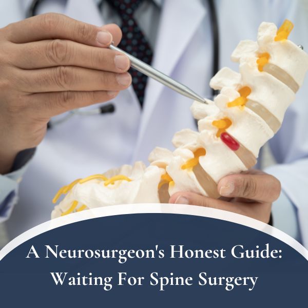
When Watching Is Wiser Than Treating
It almost always starts the same way. A patient comes in for something unrelated — a persistent headache after a car accident, dizziness that wouldn’t go away, a minor fall with a confused moment. A CT or MRI is ordered. The scan finds the original problem, which turns out to be minor. But buried in the radiologist’s report is a single sentence that will change the way this patient thinks about their health for weeks, possibly years: “Incidental finding: small enhancing lesion, likely meningioma.”
And just like that, someone who walked into the hospital for something else is now sitting across from a neurosurgeon, asking a question I hear constantly in my offices in West Islip and Smithtown: “Do I need brain surgery?”
The honest answer, most of the time, is no. At least not now. Sometimes never.
This post is the brain-tumor companion to one I wrote recently about spine surgery you probably don’t need. The theme is the same: the right surgery, at the right time, for the right patient, is one of the most powerful tools in medicine. But for many people who walk into my office with a small brain tumor on their MRI, the wisest decision is to watch and wait — not because we’re giving up, but because that’s genuinely the best medicine [1,2].
Let me explain why.
The Quiet Explosion of “Incidental” Brain Tumors
We live in the age of the MRI. Scans have gotten cheaper, faster, and more widely available. People get them for headaches, for sinus problems, after minor head bumps, as part of research studies. And as MRIs have become more common, something interesting has happened: we find a lot more small brain tumors than we used to, not because they’re becoming more common, but because we’re looking at more brains.
Most of these incidental findings are small, benign, and growing slowly — if they’re growing at all. Many have probably been there for years, quietly minding their own business, causing no symptoms and no meaningful health risk. In a landmark population study of 2,000 adults who had a research brain MRI, roughly 1.6% had a previously unknown benign primary tumor — mostly meningiomas — and the scan itself did not create the tumor [3]. It just revealed something that was already there.
That changes the calculation in a powerful way. The question stops being “how do we fix this?” and becomes “does this actually need fixing?”
The Most Common Small Brain Tumors I’m Asked About
Three types come up again and again in consultation for small, incidentally discovered brain tumors. Here’s how I think about each.
Meningiomas (the main player)
A meningioma is a tumor that grows from the meninges — the thin membranes that wrap around the brain. About 90% of meningiomas are benign (WHO Grade 1), slow-growing, and perfectly capable of being observed for years without causing harm [4].
Meningiomas are, by far, the most common tumor I’m asked to evaluate after an “incidental finding” on an MRI. They’re especially common in women, typically appear after age 50, and frequently show up in people who have no symptoms at all. Many are caught on scans done for something completely unrelated — a workup for dizziness, a concussion evaluation, even a research study.
Here’s what the data shows consistently: the majority of small, incidentally discovered meningiomas grow slowly or not at all [1,2,4]. When they do grow, the rate is usually measured i
n millimeters per year, sometimes less. Prospective studies following these tumors long-term have shown that active monitoring is safe, that growth tends to decelerate over time, and that intervention can be safely deferred — often indefinitely — in more than 40% of patients [1,2]. For a small meningioma in a person with no symptoms, surgery is frequently the wrong first move — observation with serial MRIs is often the right one.
Acoustic Neuromas (vestibular schwannomas)
An acoustic neuroma is a benign tumor that grows on the vestibulocochlear nerve — the nerve responsible for hearing and balance. These tumors are famously slow-growing. The mean growth rate is typically on the order of 0.4 to 2.9 mm per year, and a significant fraction don’t grow at all over years of follow-up [5,6]. In a Danish national cohort of more than 3,600 patients observed over 40 years, roughly 75% of tumors confined to the internal auditory canal and 60% of those extending outside it did not grow during a decade of observation [6].
Small acoustic neuromas (usually defined as under about 1.5 to 2 cm) present a textbook example of “three legitimate options”: observation, stereotactic radiosurgery, or microsurgical removal. All three can be correct answers depending on the patient, the tumor, and the trajectory. For a small tumor that isn’t causing significant hearing loss or balance problems — and isn’t clearly growing — watching is very often the right choice.
Pituitary Microadenomas
Pituitary microadenomas are a special category because the pituitary gland controls hormones. These are small tumors (under 1 cm) of the pituitary, and whether they need treatment depends heavily on whether they’re producing hormones and causing symptoms.
A “silent” microadenoma — one that’s small, not producing hormones, and causing no symptoms — often needs nothing more than periodic MRI and hormone testing. The Endocrine Society’s clinical practice guideline on pituitary incidentalomas explicitly recommends conservative follow-up for non-functioning microadenomas, with serial imaging and hormonal evaluation rather than upfront surgery [7]. A hormone-producing microadenoma, on the other hand, may absolutely need treatment — sometimes with medication rather than surgery. This is one of the clearest examples in neurosurgery of why the tum
or on the MRI is only part of the story. The clinical picture — how the patient actually feels, and what the bloodwork shows — is usually what determines the right path.
Why “Just Take It Out” Isn’t Always the Right Answer
When a patient first hears the words “brain tumor,” the instinct is almost universal: get it out of me. That instinct is understandable. It’s human. And sometimes it’s exactly right.
But brain surgery is not a small intervention. Even in the most expert hands, with the most modern technology, any operation on the brain carries real risk — infection, bleeding, stroke, neurological deficits, seizures. Those risks are generally low for modern neurosurgery, but they are not zero, and they are not trivial.
Here’s the math that gets lost in the panic of a new diagnosis: if a tumor is small, benign, growing slowly (or not at all), and causing no symptoms, the risk of surgery may be greater than the risk of the tumor itself — at least for now.
That is a very different situation from, say, a large glioma causing seizures and weakness. It’s a different situation from a meningioma pressing on the optic nerve and threatening vision. When a tumor is doing real harm, surgery is clearly the right answer and the risk-benefit math flips. But for a 7-millimeter incidental meningioma in an otherwise healthy 58-year-old? Operating right now means accepting the risks of surgery today in exchange for eliminating a risk that may never have materialized.
Sometimes that trade makes sense. Often it doesn’t.
What “Watching and Waiting” Actually Looks Like
When I recommend observation for a small brain tumor, patients sometimes hear that as “doing nothing.” It’s not. Watchful waiting is an active, structured plan — and it’s one of the most important tools in modern neurosurgery.
A typical observation protocol for a small, incidentally discovered tumor looks something like this:
- A baseline MRI with contrast. This gives us the starting point — size, location, characteristics, and anything unusual that might change the picture.
- A follow-up MRI in 6 months. The goal is to establish the tumor’s trajectory. Is it stable? Is it growing? Growing how fast?
- Annual MRIs after that, if the tumor is stable. Frequency can sometimes be reduced further after several years of confirmed stability.
- Routine clinical check-ins. Any new symptoms — headaches, vision changes, hearing loss, weakness, hormonal changes — warrant an earlier scan.
- Hormonal evaluation for pituitary tumors. Bloodwork to make sure the tumor isn’t silently overproducing or underproducing key hormones.
- A clear threshold for intervention. The patient and I agree in advance on what would change the plan — measurable growth, new symptoms, proximity to a critical structure.
That last point matters. A good observation plan isn’t vague. It has a defined trigger for when surgery or radiation moves from “maybe someday” to “yes, now.” Without that trigger, observation can drift into avoidance, which isn’t the same thing.
When I Recommend Treatment — Even for a Small Tumor
To be clear: there are situations where even a small brain tumor genuinely needs treatment. Observation is a default, not a rule. Here are the scenarios that change the conversation:
- The tumor is causing real symptoms. Seizures, progressive weakness, vision changes, significant hearing loss, or hormone problems from a pituitary tumor are all reasons to act.
- The tumor is in a dangerous location. A small tumor near the optic chiasm, the brainstem, or a critical eloquent area may need intervention earlier than a tumor in a safer location.
- The tumor is clearly growing. Confirmed, measurable growth on serial MRIs — especially accelerating growth — shifts the calculation. A tumor that’s doubled in size over two years behaves very differently from one that’s been stable for a decade.
- The imaging suggests higher-grade disease. Most meningiomas are benign, but a small percentage are atypical or malignant. Certain imaging features raise that concern and may prompt treatment even before symptoms appear.
- The patient is young with a long life ahead. A tumor that might be safely observed in an 80-year-old may warrant earlier treatment in a 45-year-old simply because the cumulative risk of growth over decades is much higher.
- The patient can’t tolerate the uncertainty of observation. This one is legitimate. Some patients do well with “watch and wait.” Others are meaningfully harmed by the ongoing anxiety, and that harm is real. A thoughtful conversation about quality of life belongs in these decisions.
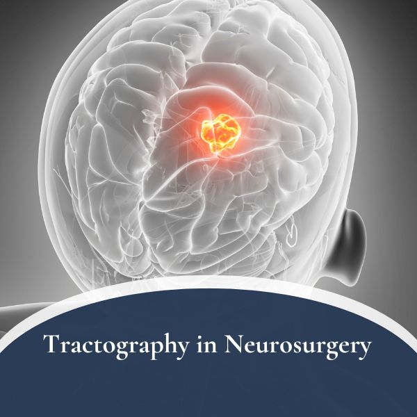
When we do treat, we have more tools than ever. Microsurgical removal remains excellent for many tumors. Stereotactic radiosurgery — essentially focused radiation delivered in one to five sessions, without cutting — has become a powerful option for small meningiomas, acoustic neuromas, and other lesions. Advanced imaging tools like tractography help us plan safer operations when surgery is the right answer. And for hormone-producing pituitary tumors, medication alone can sometimes control the disease.
The point is: having options means we can tailor the treatment to the patient, not the other way around.
A Note on Second Opinions
As with spine surgery, second opinions for a newly diagnosed brain tumor are not an insult to anyone. They’re standard good practice, especially for benign lesions where the right path isn’t always obvious.
If you’ve been told you have a small brain tumor and you’re unsure about the recommendation — whether it’s for immediate surgery or for a wait-and-see plan you don’t fully trust — seek a second opinion. Bring your actual imaging on a disc or USB drive, not just the written report. Bring your questions in writing. And look for a surgeon who will honestly tell you when watching is the right answer, not just when operating is.
The same principle applies as with my spine post: the surgeon who gives you options and honest trade-offs is the one worth listening to. That may be me. It may be someone else. Either way, you deserve that conversation.
The Bottom Line
A small brain tumor is scary. A brain tumor that doesn’t need surgery is somehow almost scarier, because it leaves patients living with a question mark in their head — literally. I understand that. But the data, the trajectory, and modern neurosurgical judgment support the same conclusion in many of these cases: for a small, benign, slow-growing tumor that isn’t causing symptoms, the best neurosurgery is often the neurosurgery we don’t do.
The right care plan isn’t always the most aggressive one. Sometimes it’s a baseline MRI, a follow-up in six months, and the reassurance that you have a plan — a real, structured, well-thought-out plan — even if that plan is “let’s keep an eye on this and make sure it doesn’t change.”
If you’ve had an incidental finding on a brain MRI and want a second opinion from a neurosurgeon who will tell you the truth — including, when it’s appropriate, the truth that you probably don’t need surgery — our Long Island offices are here to help.
Because the right surgery, at the right time, for the right patient, matters enormously. And so does the decision to wait.
Dr. Symeon Missios is a board-certified neurosurgeon practicing on Long Island, with expertise in brain and spine surgery. To schedule a consultation or second opinion, request an appointment or call (631) 422-5371 or toll-free (888) 737-5427.
References
- Strømsnes TA, et al. Growth dynamics of incidental meningiomas: a prospective long-term follow-up study. Neuro-Oncology Practice. 2023;10(3):238–248. doi:10.1093/nop/npac099
- Delgado-López PD, et al. Volumetric growth rate of incidental asymptomatic meningiomas: a single-center prospective cohort study. Acta Neurochirurgica. 2021;163(6):1665–1675. doi:10.1007/s00701-021-04815-1
- Vernooij MW, Ikram MA, Tanghe HL, et al. Incidental findings on brain MRI in the general population. New England Journal of Medicine. 2007;357(18):1821–1828. doi:10.1056/NEJMoa070972
- Islim AI, et al. Incidental intracranial meningiomas: a systematic review and meta-analysis of prognostic factors and outcomes. Journal of Neuro-Oncology. 2019;142(2):211–221. doi:10.1007/s11060-019-03110-5
- Marinelli JP, et al. Long-term natural history and patterns of sporadic vestibular schwannoma growth: a multi-institutional volumetric analysis of 952 patients. Neuro-Oncology. 2022;24(8):1298–1306. doi:10.1093/neuonc/noab303
- Reznitsky M, et al. The natural history of vestibular schwannoma growth — prospective 40-year data from an unselected national cohort. Neuro-Oncology. 2021;23(5):827–836. doi:10.1093/neuonc/noaa230
7. Freda PU, Beckers AM, Katznelson L, et al. Pituitary incidentaloma: an Endocrine Society clinical practice guideline. Journal of Clinical Endocrinology & Metabolism. 2011;96(4):894–904. doi:10.1210/jc.2010-1048
By Symeon Missios, MD — Long Island Brain & Spine