- Provides the ability to visualize paths within the subcortical white matter
- It assesses the direction and motion of water molecules within the white matter fibers
- Allows the visualization of white matter tracts in 3d and their relationships with specific objects such as a brain tumor
- It can provide information that may lead to improved extent of resection and decreased morbidity
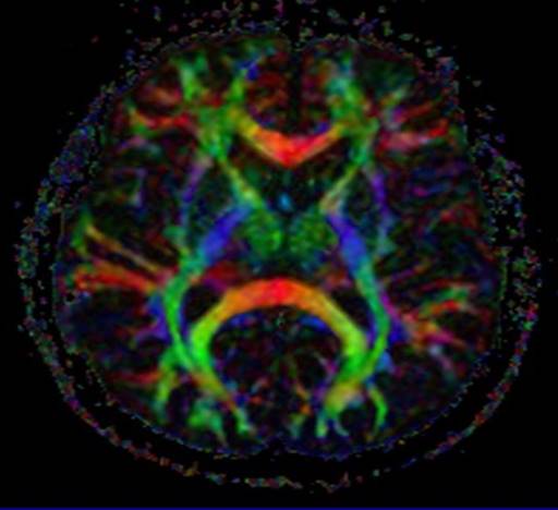
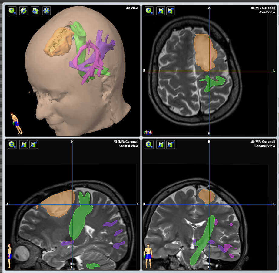
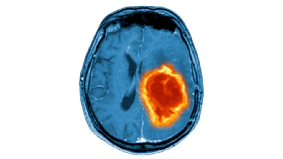
A DTI MRI reveals the anatomy and direction of the white matter fibers and assigns colors to them based on their direction of orientation. Specific fibers can be selected and converted to 3 dimensional objects which can help reveal their relationship to the tumor and help with preoperative planning as shown on the right. The video illustrates a case where a tumor (red) is very close to a fiber bundle called the arcuate fasciculus (yellow) which plays an important role in speech function.


