How Brain Tumor Surgery Is Learning to See Cancer Directly
By Symeon Missios, MD — Long Island Brain & Spine
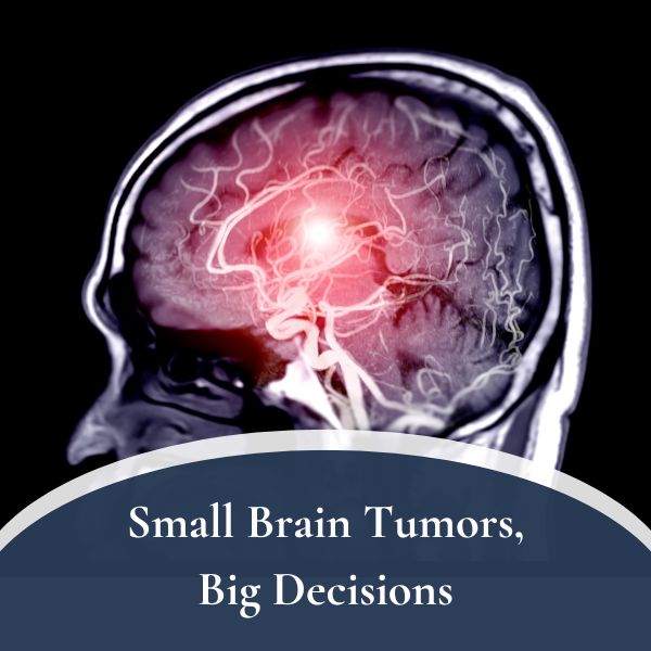
There is a moment in every brain tumor operation that never gets easier, no matter how many times you’ve stood at the table. The bulk of the tumor is out. The cavity looks clean. And you have to answer the oldest question in this work: is that enough, or is there more?
For my entire career, I’ve answered that question with my eyes and my judgment. I look at the tissue. I check it against the MRI we loaded into the navigation system before we started. I sometimes use a fluorescent dye that makes tumor cells glow pink under a special light. I weigh all of it against one unforgiving fact — that the brain tissue at the edge of a tumor is doing something a patient needs, whether that’s moving a hand, finding a word, or seeing the left side of the world.
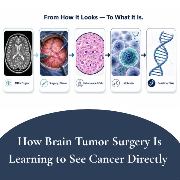
Every one of those tools is, in its own way, indirect. The MRI shows me how the tumor looked an hour ago. The dye tells me cells are probably abnormal, not how many or what kind. The microscope shows me what the tissue looks like — and at the blurry border between tumor and brain, even an expert pathologist can struggle to tell one cell from another.
That is starting to change. And the change is significant enough that I want to explain it plainly, because if you or someone you love is facing brain tumor surgery in the next few years, the way that surgery is planned and judged is about to be different from the way it has been done for the last fifty.
What “Maximal Safe Resection” Has Always Meant
If you’ve researched brain tumor surgery at all, you’ve probably run into the phrase maximal safe resection. It’s the governing principle of modern brain tumor surgery, and we owe the framework largely to the work of Dr. Mitchel Berger and others who reshaped the field. The idea is a balance. Remove as much tumor as possible, because for most tumors more removal means longer survival. But preserve the patient’s neurological function, because a “successful” operation that leaves someone unable to speak or walk is not a success.
That balance rests on two questions a surgeon has to answer in real time: Where is the tumor? and Where is the function?
We’ve gotten genuinely good at the second question. Brain mapping, awake surgery, and imaging that shows the brain’s wiring let us find and protect the parts of the brain a patient can’t live well without.
The first question — where, exactly, is the tumor — is the one we’ve actually been answering indirectly all along. And that’s the question the molecular datastream is starting to answer head-on.
What the Molecular Datastream Actually Is
Here’s the simplest way I can put it. Cancer is not, fundamentally, defined by how it looks. It’s defined by its genetics — specific mutations and chemical changes to the DNA inside the cells. A brain tumor is a tumor because of what its cells are doing at the genetic level, not because of how the tissue appears on a scan or under a microscope.
For most of medical history, we’ve had no way to read that genetic information fast enough to use it during an operation. We send tissue to the lab, and the full molecular analysis comes back days to weeks later — long after the patient has left the operating room.
The “molecular datastream” is the name for a set of new technologies that are collapsing that timeline from weeks to minutes — fast enough to actually inform what a surgeon does while the operation is still happening. Several different tools are converging here:
- Rapid imaging paired with artificial intelligence. A rapid imaging technique can now produce a microscope-quality picture of fresh tissue in the operating room in a couple of minutes, without the usual slide-preparation process. Paired with AI trained on the genetics of brain tumors, these systems can do something remarkable: predict a tumor’s genetic subtype from the image itself. One such model, developed in academic neurosurgery labs, can tell apart the two most common kinds of glioma — astrocytoma and oligodendroglioma — with accuracy approaching that of conventional pathology [1].
- Ultra-rapid genetic testing. Newer workflows can directly measure specific tumor mutations from a tissue sample in roughly fifteen minutes. By combining how many abnormal cells are present with how strongly the tumor mutation shows up, surgeons can begin to estimate the actual concentration of tumor cells at a given spot in the cavity — not infer it, measure it.
- Rapid sequencing for the full molecular fingerprint. Portable sequencing technology, again paired with AI, has been used to generate a tumor’s detailed molecular classification — one of the most precise ways we categorize brain tumors — during surgery itself, in an early proof-of-concept study of glioma patients [2].
Put those together and you get a genuinely new situation: the neurosurgeon becomes the first person to hold the tumor’s molecular identity, in the operating room, while there’s still a chance to act on it.
Why This Changes the Operation
A few specific examples of how this matters, because the abstract version doesn’t capture it.
Knowing the tumor’s subtype changes how aggressive I should be. Two gliomas that look identical under the microscope can behave completely differently. The two most common types — astrocytoma and oligodendroglioma — tend to grow at different speeds and respond differently to treatment, including, in some cases, to targeted drugs. If I know during the operation which one I’m dealing with, I can make a better decision about how hard to push at a risky edge. There are situations where, for the more favorable tumor type with good drug options, it makes sense not to chase the last bit of tumor through critical brain — because the cost (a permanent deficit) outweighs the benefit.
Knowing whether a biopsy is diagnostic, before the patient leaves. Anyone who has had a brain biopsy knows the anxiety of waiting for results. There’s also a quieter anxiety on my side of it: the worry that the small sample we took doesn’t actually contain diagnostic tissue. Molecular tools that detect the defining genetic changes can tell us, during the procedure, whether we’ve captured what we need.
Knowing when the edge is actually clean. This is the big one. Researchers measuring tumor-cell content directly at the edges of the surgical cavity have found something sobering: many edges that looked acceptable were, at the molecular level, still substantially tumor. The flip side is the promise — in some cases, surgeons could keep checking and, where it was safe, keep going until the molecular signal dropped to undetectable levels. The idea of measuring leftover tumor this way and studying how it relates to recurrence is now moving toward formal clinical trials.
The Honest Part: A Sharper Knife Is Not Automatically a Wiser One
I’ve written before, on this blog, about the spine surgery many patients don’t need and the small brain tumors that are often better watched than treated. The through-line in everything I write is the same: a more powerful tool is only as good as the judgment guiding it. The molecular datastream deserves the same honesty.
Here is the question that keeps me cautious, and it comes straight from the researchers’ own data. If a number on a screen tells me there is still tumor at a particular edge, that number will be true — and it will be pulling me to cut more, sometimes in exactly the kind of critical area, one controlling movement or speech, where my whole training says to stop. That balance has always meant weighing an invisible tumor burden against a function I can see. The molecular datastream makes the tumor burden visible and quantified. It does nothing to make the function more visible. There is a real risk that the side of the scale we can finally measure simply becomes louder than the side we still can’t — and that “operate until the signal is gone” quietly becomes a justification for taking more brain than a patient should lose.
There’s an even deeper open question. When leftover tumor cells are found at an edge and the tumor later comes back, we don’t yet know whether the cancer returned because we left those cells, or whether that region was simply the kind of place a tumor tends to regrow regardless. In other words, a perfect count of cells at the edge may still not tell us where the cancer will actually come back. The molecular signal is extraordinarily precise. Precision and meaning are not the same thing — and surgery lives in the gap between them.
None of this is an argument against the technology. It’s an argument for the same thing I always argue for: using a powerful tool in the service of judgment, not as a replacement for it.
What This Means If You’re Facing Brain Tumor Surgery Now
A few honest, practical notes:
- Most of this is emerging, not yet standard. Some of these tools are in use at major academic centers; others are in clinical trials or earlier. If you’re having surgery this year, your operation will likely still rely on the established toolkit — navigation, mapping, imaging, and experienced judgment, which remain very good.
- It’s a fair thing to ask about. If you’re being treated at or referred to an academic center, it’s reasonable to ask whether intraoperative molecular tools or rapid genetic testing are part of their program, and whether you might be a candidate for a trial.
- The molecular result matters far beyond the operating room. Identifying a tumor’s genetic profile quickly helps your whole team — it can flag a mutation that a drug can target, identify candidates for clinical trials, and shape radiation and chemotherapy decisions. The benefit isn’t only about how much tumor comes out; it’s about getting the right follow-up treatment to the right patient sooner.
The Bottom Line
For fifty years, brain tumor surgeons have answered the question “did we get it all?” using indirect clues — the way tissue looks, glows, or maps onto a scan taken before we started. We’re entering an era where we can begin to answer it directly, by reading the tumor’s own genetics in the operating room, in minutes instead of weeks. That’s one of the most exciting developments in my field, and it will, over time, make brain tumor surgery more precise and more personalized.
But precision is a means, not an end. The goal has never been to remove the most tumor — it’s to do the right thing for the patient on the table, which sometimes means removing more and sometimes means having the discipline to stop. The molecular datastream is going to give surgeons a remarkable new source of information. Whether it makes brain surgery better will depend, as it always has, on the judgment of the person holding it.
If you or a family member has been diagnosed with a brain tumor and you’d like a careful, honest conversation about the options — including which tools are genuinely ready and which are still on the horizon — our offices in West Islip and Smithtown are here to help.
Dr. Symeon Missios is a board-certified neurosurgeon practicing on Long Island, with expertise in brain and spine surgery, neurosurgical oncology, and stereotactic radiosurgery. To schedule a consultation or second opinion, request an appointment or call (631) 422-5371 or toll-free (888) 737-5427.
References
(Citations below identify the key primary sources behind the technologies described. Please verify each against PubMed before publication.)
- Hollon T, Orringer DA, et al. Artificial-intelligence-based molecular classification of diffuse gliomas using rapid, label-free optical imaging. Nature Medicine. 2023. (DeepGlioma — verify exact volume/pages.)
- Vermeulen C, et al. Ultra-fast deep-learned CNS tumour classification during surgery. Nature. 2023. (Intraoperative nanopore methylation classification, “Sturgeon” — verify exact volume/pages.)
Additional background: Sanai N, Berger MS, on glioma extent of resection and patient outcome; and the WHO Classification of Tumours of the Central Nervous System, 5th ed. (2021) for current molecular glioma classification.