A man in his late sixties is brought into the emergency room by his daughter. She tells the triage nurse she found him slumped at the kitchen table that morning, unable to lift his right arm, his speech slurred into something she could not understand. He looks at her, frightened. She looks at the clock on the wall.
She is right to look at the clock.
In acute stroke care, there is a phrase we use constantly with families, residents, and each other: time is brain. It is not a slogan. It is a clinical reality with a specific, calculable cost. A 2006 analysis estimated that during a typical large-vessel ischemic stroke, the brain loses approximately 1.9 million neurons per minute that blood flow is not restored, along with roughly 14 billion synapses and 7.5 miles of myelinated nerve fibers [1]. Every hour without treatment ages the affected brain by roughly 3.6 years.
Most patients and families do not see what happens between the 911 call and the moment someone walks back out of the hospital — or does not. Here is what that window actually looks like, why every step is built around saving minutes, and why the choice of where to take a stroke patient matters as much as how fast you call.
Stroke Is a Blocked Pipe, Not a Mystery
Roughly 87% of strokes are ischemic — caused by a clot blocking blood flow to part of the brain [2]. The rest are hemorrhagic — bleeding either within the brain itself or around it. The two are treated almost oppositely, which is one reason imaging happens fast: we cannot give a clot-dissolving drug to someone whose problem is bleeding.
In an ischemic stroke, the affected brain tissue exists in two zones. The core is the tissue that has already died and cannot be saved. Around it is the penumbra — tissue that is starving but still alive, still salvageable if blood flow is restored quickly. The entire goal of acute stroke treatment is to rescue the penumbra before it joins the core. Every minute that passes, more penumbra becomes core.
Recognizing It at Home: BE FAST
The single most important variable in stroke outcomes is how quickly someone gets to a hospital — and that depends on someone in the room recognizing what is happening. The mnemonic the American Stroke Association recommends is BE FAST:
- Balance — sudden loss of balance or coordination
- Eyes — sudden vision change or trouble seeing in one or both eyes
- Face — drooping on one side; ask the person to smile
- Arm — weakness in one arm; ask them to raise both arms and see if one drifts down
- Speech — slurred, garbled, or absent speech; ask them to repeat a simple sentence
- Time — call 911 immediately and note when symptoms started
The time piece is often misunderstood. We do not need an exact start time, but we do need to know the last moment the person was clearly normal. If someone wakes up with symptoms, the last seen well time is when they went to bed — which has major implications for what treatments are still on the table.
The other piece families sometimes miss: do not drive the person to the hospital yourself. Call 911. Paramedics begin treatment in the ambulance, alert the hospital before arrival, and route the patient to a facility equipped for stroke care. A self-driven trip to the wrong hospital can cost hours that the brain does not have.
The First Sixty Minutes at the Hospital
When an EMS team calls in a “stroke alert,” the receiving hospital triggers a sequence designed to compress what used to take half a day into about forty-five minutes:
- A neurologist and a stroke team meet the ambulance at the door
- The patient goes directly to CT — usually skipping the waiting room entirely
- A non-contrast CT scan rules out bleeding
- A CT angiogram (CTA) of the head and neck looks for the location of the blockage
- Often, a CT perfusion study estimates how much penumbra is still salvageable
- Bloodwork, an EKG, and a focused neurological exam happen in parallel
By around the forty-five-minute mark, the team has enough information to make two decisions: is this patient a candidate for clot-dissolving medication, and is this patient a candidate for mechanical thrombectomy?
Clot-Busting Medication: tPA and Tenecteplase
For most ischemic strokes presenting within 4.5 hours of symptom onset, intravenous alteplase (tPA) or, increasingly, tenecteplase is the standard first-line treatment [3]. These are clot-dissolving drugs given through an IV that work systemically to break up the blockage.
The trade-off is that they thin the blood, which raises the small but real risk of bleeding — including bleeding inside the brain. That risk is the reason the criteria for who can receive these drugs are strict, the reason the CT scan happens first, and the reason patients with very small or very large strokes, very high blood pressure, or recent surgery may not be candidates.
For the right patients in the right window, IV thrombolysis can dramatically improve outcomes. But for the largest, most disabling strokes — strokes caused by clots in the major arteries at the base of the brain — IV medication alone is often not enough. That is where mechanical thrombectomy comes in.
Mechanical Thrombectomy: Pulling the Clot Out
For patients with a large vessel occlusion — a clot blocking a major artery like the internal carotid or middle cerebral — the most important treatment is no longer a drug. It is a catheter.
Mechanical thrombectomy is an endovascular procedure: a thin catheter is threaded from the groin (or sometimes the wrist) up through the vasculature to the clot in the brain. A specialized device — a stent retriever or aspiration catheter — grabs or vacuums the clot, and the artery reopens. From skin puncture to clot removal, this can take less than thirty minutes in an experienced center.
The data here are striking. A 2016 meta-analysis of five randomized trials (the HERMES collaboration) showed that thrombectomy roughly doubled the chance of patients being functionally independent at 90 days, compared with medication alone [4]. The number-needed-to-treat for one additional patient to be independent was 2.6 — among the most powerful effect sizes in modern medicine.
Equally important, the treatment window keeps expanding. The DAWN and DEFUSE 3 trials extended the eligibility window for thrombectomy out to 24 hours from last known well in carefully selected patients whose imaging shows a small core and a large salvageable penumbra [5,6]. That means even patients who wake up with stroke symptoms — historically considered “too late” — may still be treatable.
But the time-dependence within that window is brutal. Every hour of delay reduces the odds of a good outcome. “Late” is not the same as “too late,” but earlier is still always better.
Why Comprehensive Stroke Centers Matter
Not every hospital can deliver thrombectomy. The procedure requires a 24/7 team including a vascular neurosurgeon or neuro-interventionalist, neurointensivists, neuroradiologists, advanced imaging, and a dedicated neuro-ICU. Hospitals certified at this level are called Comprehensive Stroke Centers — the highest of three certification tiers, alongside Primary Stroke Centers and Thrombectomy-Capable Centers.
Good Samaritan University Hospital is part of the Stroke and Brain Aneurysm Center of Excellence at Catholic Health and is among the comprehensive stroke programs serving Long Island. When EMS suspects a large vessel occlusion, they can bypass closer hospitals to bring the patient to a center equipped to do thrombectomy — a decision that saves lives. For more on regional stroke care, see strokecarelongisland.com.
For patients and families on Long Island, this is worth knowing in advance, not in the middle of a crisis. The decision of where to go for stroke care should be made before the stroke happens, not at the hospital door.
What Families Should Know in the First Twenty-Four Hours
If a loved one is being treated for acute stroke, here is what to expect:
- They will be admitted to a neuro-ICU or stroke unit, not a general medical floor
- Blood pressure will be tightly controlled — sometimes intentionally kept higher than usual to perfuse threatened tissue
- A repeat CT or MRI at 24 hours is standard to look for completed infarct or bleeding complications
- A swallow evaluation is mandatory before any food or water — stroke commonly affects swallowing, and aspirating into the lungs is a common, preventable cause of post-stroke pneumonia
- A search for the cause of the stroke begins immediately: echocardiogram, heart rhythm monitoring, carotid imaging, bloodwork for clotting disorders
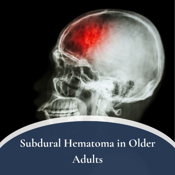
Most strokes are not strictly random. Finding the underlying cause is how we prevent the next one — which is often more damaging than the first. In older patients, a related concern worth understanding is subdural hematoma, the “slow” brain bleed, which can present similarly to stroke and is sometimes part of the differential.
What I Tell Families
Two things, almost always.
First: the first day is the worst day for predictions. Stroke patients commonly look much worse on day one than they will on day five or day thirty. The brain that has been deprived of blood swells, and the swelling itself causes symptoms that resolve as it settles. Resist the urge to make decisions about long-term function during the acute window.
Second: rehabilitation is treatment, not consolation. Aggressive, early, intensive physical therapy, occupational therapy, and speech therapy in the weeks after stroke are not optional add-ons. They are how the brain reorganizes around the injury. Patients who get into a structured rehabilitation program early do measurably better than those who do not.
The Bottom Line
Stroke is not a mystery and not a death sentence. It is a treatable, time-dependent medical emergency in which the brain is asking, by the minute, for the blockage to be cleared. The single most powerful intervention is to recognize it, call 911, and get to the right kind of hospital — not the closest, the right one.
For patients on Long Island, this is one of the few medical situations where knowing the system in advance can change the outcome. If you or a family member has had a stroke, a TIA, or a serious risk factor like atrial fibrillation, carotid disease, or a known brain aneurysm, the time to have the conversation is now, not after the alarm goes off.
Our offices in West Islip and Smithtown are here for evaluation, second opinions, and longer-term planning for patients with cerebrovascular disease. For acute symptoms — face drooping, arm weakness, slurred speech, sudden vision or balance change — do not call the office. Call 911.
Dr. Symeon Missios is a board-certified neurosurgeon practicing on Long Island, with expertise in brain and spine surgery, cerebrovascular disease, and stereotactic radiosurgery. To schedule a consultation or second opinion, request an appointment or call (631) 422-5371 or toll-free (888) 737-5427.
References
- Saver JL. Time is brain—quantified. Stroke. 2006;37(1):263–266. doi:10.1161/01.STR.0000196957.55928.ab
- Tsao CW, Aday AW, Almarzooq ZI, et al. Heart Disease and Stroke Statistics—2023 Update: A Report From the American Heart Association. Circulation. 2023;147(8):e93–e621. doi:10.1161/CIR.0000000000001123
- Powers WJ, Rabinstein AA, Ackerson T, et al. Guidelines for the Early Management of Patients With Acute Ischemic Stroke: 2019 Update. Stroke. 2019;50(12):e344–e418. doi:10.1161/STR.0000000000000211
- Goyal M, Menon BK, van Zwam WH, et al. Endovascular thrombectomy after large-vessel ischaemic stroke: a meta-analysis of individual patient data from five randomised trials. Lancet. 2016;387(10029):1723–1731. doi:10.1016/S0140-6736(16)00163-X
- Nogueira RG, Jadhav AP, Haussen DC, et al. Thrombectomy 6 to 24 Hours after Stroke with a Mismatch Between Deficit and Infarct. N Engl J Med. 2018;378(1):11–21. doi:10.1056/NEJMoa1706442
- Albers GW, Marks MP, Kemp S, et al. Thrombectomy for Stroke at 6 to 16 Hours with Selection by Perfusion Imaging. N Engl J Med. 2018;378(8):708–718. doi:10.1056/NEJMoa1713973
By Symeon Missios, MD — Long Island Brain & Spine