A “Slow” Brain Bleed (A Neurosurgeon’s Friendly Guide)
If you spend time in any neurosurgery clinic—or with a loved one in their 60s, 70s, or beyond—you’ll hear the phrase “subdural hematoma” more often than you’d expect. It sounds dramatic, and it can be. But it’s also a condition we treat every week with excellent outcomes when it’s recognized in time.
Think of a subdural hematoma (SDH) as blood collecting in a thin space between the brain and its covering. In older adults, it often doesn’t gush; it oozes—slowly, almost politely—after what felt like a nothingburger of a fall or bump. Days to weeks later, that small ooze adds up. The pressure builds. And a person who “just felt off” starts to shuffle, forget words, or lean to one side. That slow pace is why doctors sometimes call it a “slow brain bleed.”
Why it happens (and why age matters)
As we age, the brain naturally shrinks a bit—it’s normal. That gentle shrinkage stretches the tiny bridging veins that cross from brain to covering. A minor fall, a quick jolt in the car, even an overzealous head scratcher (I’m only half kidding) can tear one of these veins. If you’re taking blood thinners (like warfarin, DOACs) or antiplatelets (like aspirin, clopidogrel), the risk of persistent bleeding rises. The result is often a chronic subdural hematoma: blood that collects slowly, sometimes mixes with fluid, and irritates the membrane so it keeps seeping.
Not every subdural is slow. A fresh, acute subdural can appear after a major head injury and cause rapid decline. But in older adults living independently, it’s the chronic, gradual variety we encounter most.
Who’s at higher risk?
- Adults >60–65 (more brain “slack,” more fragile veins)
- Blood thinners/antiplatelets (warfarin, DOACs, aspirin, clopidogrel)
- Frequent falls or balance issues
- Alcohol use disorder (nutritional factors, falls)
- Prior subdural hematoma or recent head trauma
How it shows up (it’s not always a headache)
Headache plays a role, but it’s not the whole story. What families describe is subtler—and that’s the danger. I’ll often hear, “He’s just not himself.” We see slowed thinking, memory slips, and personality change. Many patients develop a worsening shuffle or drifting to one side, weakness in a hand or leg, or frequent falls that seem to come out of nowhere. Sometimes there’s speech difficulty or a new seizure. A few notice double vision. The onset can be so gradual that the day it “really started” is hard to pin down.
Red flags that deserve same-day evaluation
- A new or worsening headache after a fall (even a “small” one)
- Confusion, sleepiness, or personality change
- Weakness, numbness, or clumsiness—often on one side
- Trouble walking, shuffling, or new frequent falls
- Slurred speech, difficulty finding words, or facial droop
- Seizure (first-time seizure always needs a look)
- Any of the above in someone on blood thinners/antiplatelets
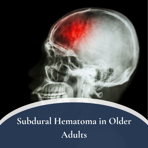
What happens in the ER (and why CT is king)
Evaluation starts with a story: what happened, when, and what changed since. Then a focused neurological exam—strength, sensation, balance, eye movements, speech. The test that answers the big question fastest is a CT scan of the head. It’s quick, widely available, and excellent at spotting blood.
A chronic subdural often looks like a crescent hugging the brain. It may be on one side or both. It may look darker (older blood) or have mixed shades if there have been re-bleeds. The amount of shift—how much the brain is pushed—matters as much as the size. An MRI can add detail in borderline cases, but most decisions are made right off the CT.
Do all subdural hematomas need surgery?
No. Small, minimally symptomatic chronic subdurals can sometimes be observed with close follow-up and repeat imaging. If symptoms are mild and the collection isn’t causing pressure or shift, watching is reasonable. But many patients do benefit from a procedure, and the aim is simple: create space for the brain by removing the collection and stop the cycle that refills it.
Treatment options at a glance
- Watchful waiting: for small, minimally symptomatic collections; frequent re-checks.
- Surgery to drain the hematoma: most often burr holes (tiny openings to wash out fluid and place a short-term drain). Some cases need a mini-craniotomy (a larger window) if the blood is thick or compartmentalized.
- MMA embolization (middle meningeal artery embolization): a catheter-based procedure to shut down the tiny vessels feeding the hematoma membranes—used as an adjunct to surgery or, in selected cases, as a standalone treatment.
- Medication optimization: manage blood thinners/antiplatelets safely, treat seizures if present, and prevent blood pressure spikes.
Let’s unpack the two procedures families ask me about most: burr hole drainage and MMA embolization.
Burr hole surgery: the classic, effective workhorse
Despite the high-tech era, the most common and effective first step for many symptomatic chronic subdurals is still burr hole drainage. Under anesthesia, we make one or two small openings in the skull right over the collection. We gently irrigate the subdural space to wash out liquefied blood, then place a soft drain for 1–2 days to keep fluid from reaccumulating as the brain re-expands. Most patients notice improvement within hours to a day—clearer thinking, steadier walking, a lighter head.
Complications are uncommon but can include infection, bleeding, or recurrence (the hematoma coming back). Recurrence happens because the inner membrane may still ooze; even after a great washout, that membrane sometimes needs extra help to quiet down. That’s where MMA embolization enters the conversation.
MMA embolization: cutting the supply lines to prevent recurrences
Middle meningeal artery (MMA) embolization is a minimally invasive procedure done by an endovascular specialist. Through a tiny artery puncture in the wrist or groin, we navigate a micro-catheter up to the middle meningeal artery, which nourishes the membrane that lines the hematoma. We then inject particles or liquid embolic to seal off those tiny feeders. Think of it as turning off the faucet that keeps the subdural space damp and prone to refilling.
How we use it:
- Adjunct to burr holes: drain now for fast symptom relief, and embolize to reduce recurrence risk.
- Primary therapy in selected patients: for patients with mild-to-moderate symptoms, high anesthesia risk, or recurrent subdurals, MMA embolization may be offered without immediate surgery. It doesn’t relieve pressure as fast as drainage, so we choose carefully based on symptoms and imaging.
What to expect:
- Most procedures are same-day or overnight stays.
- Small puncture site; walking within hours.
- Benefits accrue over weeks as the membrane quiets and the collection resorbs.
- Risks are low but include vessel injury, stroke, or non-target embolization—uncommon in experienced hands.
Families often ask, “Which is better, burr holes or embolization?” The honest answer is they do different jobs. Burr holes decompress now; embolization discourages recurrence. For many patients, both is the winning combination—relief today and fewer comebacks tomorrow.
What recovery looks like
After burr holes, patients typically spend 1–2 days in the hospital with a drain and repeat imaging. We encourage early mobility, physical therapy, and careful attention to blood pressure, hydration, and medication management. Some patients feel dramatically clearer right away; others improve over days to weeks as the brain re-expands. If MMA embolization is part of the plan, it may occur during the same admission or scheduled soon after discharge.
We’ll also talk about driving, fall precautions, and when to resume blood thinners if those are essential (for a heart valve, atrial fibrillation, etc.). That decision is individualized, balancing bleed risk against clot risk, and often involves your cardiologist or primary doctor.
Can it come back?
Yes—recurrence is the challenge with chronic subdurals. Even after a clean drainage, the irritated membrane can re-seep. That’s why we see you in clinic, repeat a scan, and keep a close eye on symptoms. MMA embolization has emerged as a valuable recurrence-reducer in many centers. Not every patient needs it, but for bilateral hematomas, very large collections, anticoagulation, or a prior recurrence, I discuss it early.
Practical questions families ask me
“Grandma fell a month ago and only now seems off—could this still be related?”
Yes. A chronic subdural commonly declares itself days to weeks after seemingly minor trauma.
“She’s on a blood thinner. Is surgery still safe?”
We operate on blood-thinner patients frequently. We’ll reverse or hold the medication safely if possible and coordinate re-starting it later. The key is individualized risk balancing.
“If we do embolization alone, will she feel better right away?”
Embolization doesn’t decompress like drainage; it prevents re-bleeding and speeds resorption. If symptoms are significant, we often drain first, then embolize.
“Does a subdural hematoma mean dementia?”
No. Subdural can mimic dementia—slowed thinking, memory changes—but many patients sharpen right up after treatment.
Prevention (or at least, stacking the deck)
While you can’t bubble-wrap real life, you can lower risk:
- Review medications: make sure blood thinners/antiplatelets are truly needed and dosed correctly; avoid unnecessary duplications (e.g., aspirin + clopidogrel without a reason).
- Fall-proof the home: good lighting, remove loose rugs/clutter, grab bars in bath, non-slip shoes, clear pathways.
- Strength & balance: simple PT exercises, tai chi or chair yoga, and a daily walking habit matter.
- Vision & footwear: update glasses; shoes that grip and fit.
- Alcohol moderation: it reduces falls and improves nutrition.
When to call, when to wait
- Call now / go to the ER if there’s worsening confusion, new weakness, slurred speech, repeated falls, severe headache after a head bump, or seizure, especially in someone on blood thinners.
- Call soon (days) if there’s a subtle but persistent change—gait slowing, forgetfulness, one-sided clumsiness—even without headache, particularly after a recent fall.
- Reasonable to watch only applies when symptoms are minimal and a clinician has already seen the scan and set a follow-up plan.
Bottom line
A subdural hematoma in older adults is common, often treatable, and sometimes curable—but it hides in plain sight. If a loved one seems “a little off” weeks after a bump, respect that instinct. Diagnosis is straightforward with a CT scan. Treatment ranges from watchful waiting to burr hole drainage and, increasingly, middle meningeal artery (MMA) embolization to cut the supply lines and reduce recurrences. The best outcomes come from early recognition, individualized planning, and a team that talks to each other—and to you.
Friendly disclaimer: This article is general education, not personal medical advice. If your symptoms match the red flags above—or you’re worried—please seek care promptly.