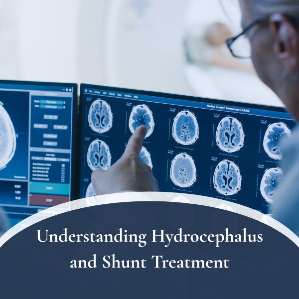
Hydrocephalus, a condition characterized by abnormal cerebrospinal fluid (CSF) accumulation within the brain’s ventricles, is often associated with infants and children. However, it’s essential to recognize that adults can also develop this condition, albeit less frequently. Dr. Symeon Missios, a renowned neurosurgeon specializing in hydrocephalus treatment in St. James, has identified five crucial signs that adults should be mindful of, as early detection is paramount for effective management and improved outcomes.
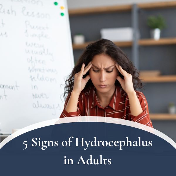
Persistent Headaches:
One of the hallmark symptoms of hydrocephalus in adults is persistent headaches. It is commonly described as dull, throbbing, or pressure-like and may worsen upon waking in the morning or with activities that increase intracranial pressure, such as coughing or sneezing. Dr. Missios emphasizes the importance of not dismissing these headaches as mere stress or tension headaches without a thorough medical evaluation, as they could indicate underlying hydrocephalus.
Cognitive Changes:
Hydrocephalus can significantly impact cognitive function in adults. Memory loss, difficulty concentrating, and decreased mental sharpness are common cognitive changes associated with the condition. Patients may notice difficulties in performing daily tasks, maintaining focus, or processing information, which should prompt them to seek medical attention promptly.
Visual Problems:
Visual problems, such as blurred vision, double vision (diplopia), or difficulty focusing the eyes, can happen because of pressure on the optic nerves caused by hydrocephalus. Dr. Missios underscores the significance of promptly addressing visual changes to prevent permanent damage to the optic nerves and preserve vision.
Balance and Coordination For Hydrocephalus Treatment in St James:
Hydrocephalus can disrupt the brain’s normal functioning, leading to balance and coordination problems in adults. Gait disturbances, unsteadiness, and difficulty maintaining balance may occur, significantly impacting an individual’s mobility and quality of life if left untreated.
Urinary Incontinence:
Changes in urinary habits, such as increased frequency, urgency, or incontinence, can indicate hydrocephalus affecting the brain regions responsible for controlling bladder function. Dr. Missios stresses the importance of discussing urinary changes openly with a healthcare provider to ensure appropriate evaluation and management.
Contact Us For Hydrocephalus Treatment in St James
Recognizing these signs of hydrocephalus in adults is crucial for timely diagnosis and treatment. Dr. Symeon Missios, MD, offers comprehensive hydrocephalus treatment in St. James, prioritizing patient care and outcomes. If you or a loved one experience any of these symptoms, it’s essential to seek medical attention promptly to ensure the best possible prognosis and quality of life.