Meningiomas are a common type of brain tumor that many people might not be aware of. As a neurosurgeon, I frequently encounter patients with questions about meningiomas, their symptoms, and treatment options. In this comprehensive guide, I will provide an overview of everything you need to know about meningiomas, from their nature and presenting symptoms to diagnosis and treatment options.
What are Meningiomas?
Meningiomas are typically benign (non-cancerous) tumors that arise from the meninges, the protective layers covering the brain and spinal cord. They account for approximately 30% of all primary brain tumors and can occur at any age but are more common in middle-aged and older individuals, with a higher prevalence in women. Although most meningiomas are benign, they can cause various symptoms depending on their location, size, and growth rate. Some meningiomas may become malignant (cancerous), but this is rare.
Types of Meningiomas
Meningiomas are classified based on their microscopic appearance and growth patterns. The World Health Organization (WHO) has divided meningiomas into three grades:
- Grade I: Benign meningiomas, which are slow-growing and have a low risk of recurrence after treatment.
- Grade II: Atypical meningiomas, which grow more aggressively and have a higher risk of recurrence.
- Grade III: Malignant or anaplastic meningiomas, which are fast-growing, invasive, and have a high risk of recurrence and spreading to other parts of the body.
Risk Factors and Causes
The exact cause of meningiomas remains unclear, but several factors are thought to increase the risk of developing these tumors. Some of the identified risk factors include:
- Age: The risk of meningiomas increases with age, with most cases occurring in people between 40 and 70 years old.
- Gender: Women are more likely to develop meningiomas than men.
- Radiation exposure: Exposure to ionizing radiation, such as radiation therapy for head and neck cancers, increases the risk of meningiomas.
- Genetic factors: Certain genetic conditions, such as neurofibromatosis type 2, are associated with an increased risk of meningiomas.
Presenting Symptoms
The symptoms of meningiomas can vary depending on the tumor’s location within the brain or spinal cord. Some common presenting symptoms include:
- Headaches
- Seizures
- Weakness or numbness in the limbs
- Visual disturbances
- Memory and concentration problems
- Personality or mood changes
- Balance and coordination issues
It is essential to note that these symptoms can also be caused by many other conditions, so proper evaluation and diagnosis by a medical professional are necessary.
Diagnosis
If a meningioma is suspected, your doctor will likely recommend one or more imaging tests to confirm the diagnosis and gather information about the tumor’s size, location, and growth rate. These tests may include:
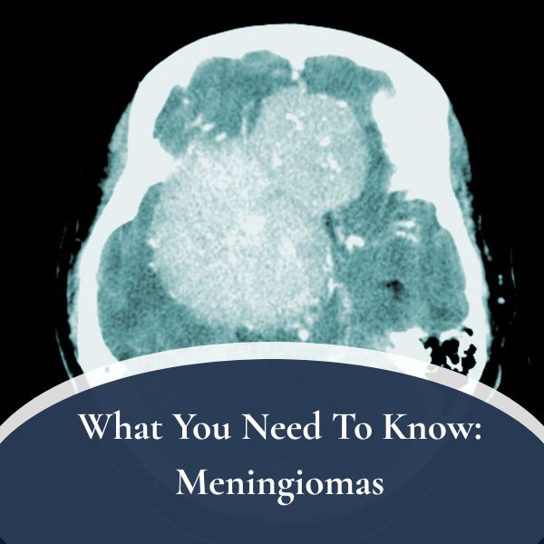
4.1. CT Scan
A computed tomography (CT) scan is an X-ray technique that generates detailed cross-sectional images of the brain. It can help identify meningiomas and determine their size and location. Sometimes, a contrast material is injected into the bloodstream to enhance the visibility of the tumor.
4.2. MRI
Magnetic resonance imaging (MRI) uses a magnetic field and radio waves to generate detailed images of the brain and spinal cord. It is often the preferred imaging method for diagnosing meningiomas, as it provides better soft tissue contrast and allows for more precise identification of tumor location, size, and nearby structures.
Treatment Options
The treatment of meningiomas depends on several factors, including the tumor’s size, location, growth rate, and the patient’s overall health. There are three primary treatment options for meningiomas:
5.1. Observation and Serial Imaging
For small, asymptomatic meningiomas with slow growth rates, a “watchful waiting” approach may be recommended. In this case, the patient will undergo regular imaging tests (CT or MRI) to monitor the tumor’s growth and development. If the tumor begins to grow or cause symptoms, more aggressive treatment options may be considered.
5.2. Stereotactic Radiosurgery
Stereotactic radiosurgery is a non-invasive treatment that delivers a high dose of radiation directly to the tumor while sparing the surrounding healthy tissue. It is typically used for small to medium-sized meningiomas and can be a suitable option for patients who are not good candidates for surgery due to their health or tumor location. Stereotactic radiosurgery is often performed using advanced technologies like the Varian LINAC, the Gamma Knife or CyberKnife platforms.
5.3. Surgical Resection
Surgical resection, or the removal of the tumor, is the most common treatment for meningiomas. The goal is to remove as much of the tumor as possible while minimizing damage to surrounding brain tissue. In some cases, complete removal of the tumor is possible, while in others, only a portion of the tumor can be safely removed. The type of surgery will depend on the tumor’s size, location, and proximity to critical brain structures.
Postoperative Care and Recovery
Following meningioma treatment, postoperative care and recovery will vary depending on the chosen treatment method and the patient’s individual needs. For patients who undergo surgical resection, postoperative care typically includes:
- Pain management: Medications will be prescribed to manage pain and discomfort after surgery.
- Monitoring: Your medical team will closely monitor your vital signs, neurological function, and potential complications.
- Physical therapy: Rehabilitation may be recommended to help restore strength, mobility, and balance.
- Follow-up imaging: Regular imaging tests (CT or MRI) will be scheduled to monitor for any signs of tumor recurrence.
For patients who undergo stereotactic radiosurgery, post-treatment care may include:
- Monitoring: Your medical team will monitor your response to the treatment and any potential side effects.
- Follow-up imaging: Regular imaging tests will be scheduled to assess the tumor’s response to the radiation and monitor for any signs of recurrence. It is essential to follow your medical team’s instructions and attend all follow-up appointments to ensure the best possible outcome.
Schedule With Dr. Symeon Missios
Meningiomas are common brain tumors that can cause a range of symptoms depending on their size and location. Diagnosis typically involves imaging tests like CT scans and MRIs. Treatment options include observation and serial imaging, stereotactic radiosurgery, or surgical resection, depending on the patient’s specific needs. Postoperative care and recovery will vary based on the chosen treatment method and individual circumstances. If you suspect you may have a meningioma or have questions about your condition, consult with a medical professional to discuss your concerns and determine the best course of action for your situation. Contact us today for more!